





Background: Sleep Regulation and the Subcortical Paradigm
For decades, the dominant model of sleep control placed the brainstem, hypothalamus, and thalamus at the helm. These subcortical structures generate the rhythmic firing patterns that cascade upward, imposing the characteristic slow‑wave activity on the cortex. The cortex, in turn, was viewed as a passive recipient—its neurons synchronizing to an external pacemaker rather than originating the rhythm themselves.
Key concepts that have shaped this view include:
- Ventrolateral preoptic nucleus (VLPO) – a hypothalamic hub that releases GABA and galanin to silence wake‑promoting nuclei.
- Ascending arousal system – cholinergic, monoaminergic, and orexinergic pathways that keep the cortex alert.
- Slow‑wave sleep (SWS) – large‑amplitude, low‑frequency oscillations that appear first in the cortex but were thought to be driven by thalamic relay neurons.
The new study challenges every bullet point above by demonstrating that a cortical microcircuit can independently initiate the sleep state.
The Landmark Nature Study: Methods and Findings
The research team, led by Geoffrey Terral and Renata Batista‑Brito at Albert Einstein College of Medicine, published their findings in Nature. Their experimental pipeline combined cutting‑edge genetics, optogenetics, and in‑vivo electrophysiology.
Core discoveries
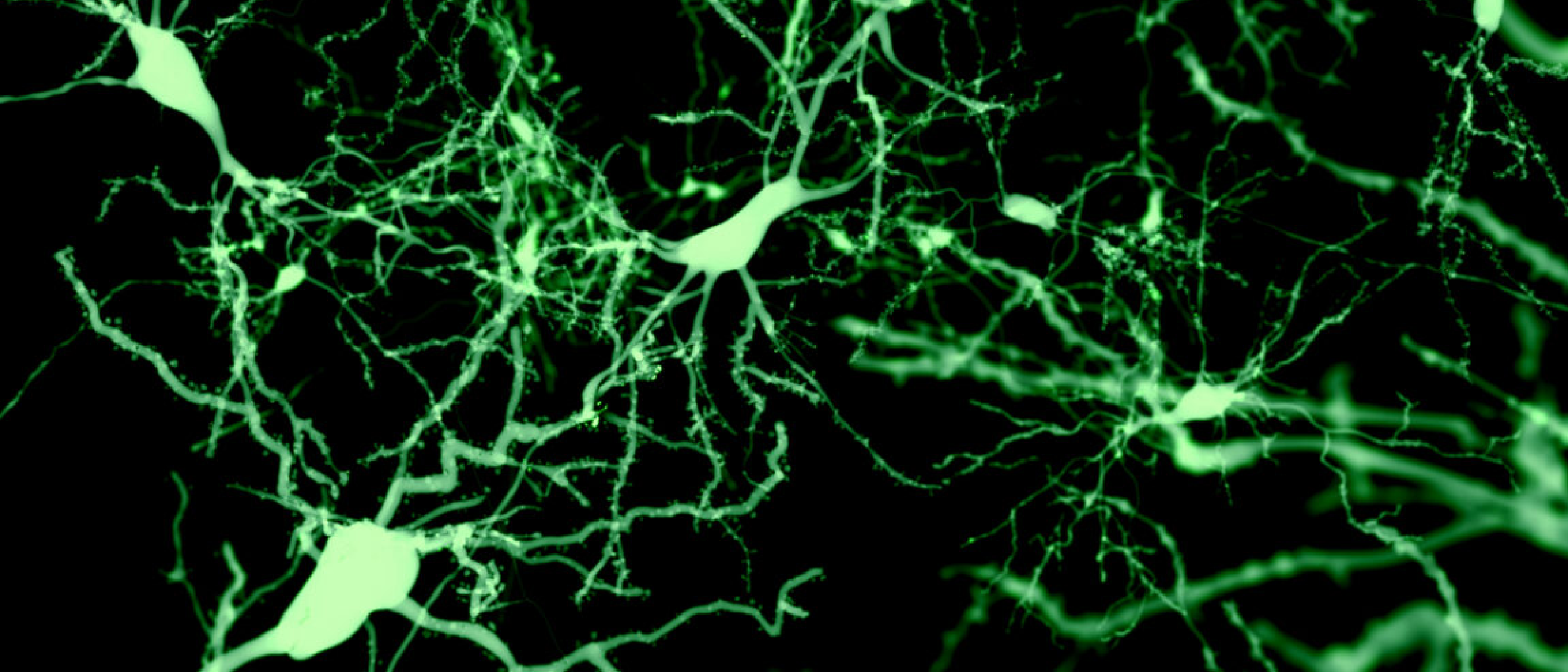
- Identification of a distinct inhibitory population – Roughly 1 % of all cortical inhibitory neurons expressed a unique molecular signature (e.g., somatostatin‑positive, neuropeptide Y‑co‑expressing).
- Causal activation induces sleep – Using Cre‑dependent channelrhodopsin‑2 (ChR2) expressed selectively in these cells, the team delivered blue‑light pulses to the mouse cortex. Within seconds, the animals entered a stable NREM‑like state, confirmed by EEG slow‑wave patterns and behavioral immobility.
- Sufficiency and necessity – Silencing the same cells with halorhodopsin prevented the natural onset of sleep during the dark phase, indicating that these neurons are not merely permissive but essential for sleep initiation.
“Usually, sleep is associated with being controlled by subcortical regions,” said Geoffrey Terral.
“What our work shows is that the cortex can not only see this rhythm but also initiate it by itself, and this is sufficient to promote sleep,” Terral added.
Experimental rigor
- Cell‑type specificity was validated through single‑cell RNA sequencing, confirming the molecular fingerprint of the targeted population.
- Temporal precision: Light pulses of 10 ms at 20 Hz were sufficient; longer stimulation did not further deepen sleep, suggesting a threshold effect.
- Cross‑validation: Similar results were reproduced in both male and female mice, and in two distinct cortical regions (prefrontal and somatosensory), underscoring the generality of the mechanism.
Why It Matters: Scientific and Clinical Implications
Redefining sleep neurobiology
The discovery forces a revision of the hierarchical model of sleep regulation. Rather than a one‑way command from subcortical nuclei, the cortex now appears to possess bidirectional control, capable of both sensing and generating the slow‑wave rhythm. This aligns with emerging theories that view the brain as a distributed network of oscillators that can self‑organize.
Therapeutic potential
- Insomnia treatment – Pharmacological agents that selectively enhance the activity of this cortical inhibitory subset could provide a novel class of hypnotics with fewer side effects than current GABA‑ergic drugs.
- Neurodegenerative disease – Slow‑wave sleep is critical for glymphatic clearance of metabolic waste. Boosting cortical‑driven sleep may slow the accumulation of amyloid‑β and tau, offering a non‑invasive adjunct for Alzheimer’s disease.
- Precision neuromodulation – Non‑invasive brain stimulation (e.g., transcranial magnetic stimulation) could be tuned to the identified frequency and spatial pattern to activate these cells without surgery.
Broader tech‑health crossover
The intersection of biology and technology highlighted by this study mirrors concerns raised in the AI‑Designed Viruses article, where synthetic biology tools can both cure and create. Understanding how to safely manipulate neuronal circuits is essential to avoid unintended consequences, much like the bio‑security discussions surrounding engineered pathogens.
Technical Breakdown: How Cortical Inhibitory Neurons Were Manipulated
Genetic targeting strategy
- Cre‑driver line – Researchers used a mouse line expressing Cre recombinase under the promoter of a gene uniquely active in the identified inhibitory subset.
- AAV‑mediated opsin delivery – An adeno‑associated virus (AAV) carrying a double‑floxed inverted ChR2‑eYFP cassette was injected stereotaxically into the cortex. Cre recombination flipped the cassette into the correct orientation, restricting expression to the target cells.
Optogenetic stimulation protocol
- Fiber‑optic implantation – Thin optic fibers were positioned 200 µm above the injection site to minimize tissue damage.
- Stimulation parameters – 10 ms pulses at 20 Hz for 30 seconds reliably induced sleep; longer trains did not increase depth, indicating a binary switch rather than a graded response.
Electrophysiological verification
- EEG/EMG recordings – Simultaneous cortical EEG and neck EMG confirmed the transition from wakeful desynchronized activity to high‑amplitude, low‑frequency delta waves.
- Local field potentials (LFPs) – LFPs recorded from the stimulated region showed a rapid increase in delta power within 2 seconds of light onset.
Safety and translational considerations
- Off‑target effects – Whole‑brain c‑Fos mapping revealed minimal activation outside the targeted cortical area, reducing concerns about widespread inhibition.
- Potential for human translation – While optogenetics is not yet clinically viable, chemogenetic approaches (e.g., DREADDs) or **ultrasound‑mediated neuromod
‑mediated neuromodulation could bypass the need for invasive implants, allowing clinicians to activate the sleep‑inducing circuit through focused acoustic energy that crosses the skull. Early pilot studies in rodents have shown that low‑intensity transcranial ultrasound can selectively depolarize genetically sensitized neurons, offering a plausible pathway toward non‑invasive human therapies.
Roadblocks on the path to bedside
| Challenge | Why it matters | Possible mitigation |
|---|---|---|
| Cell‑type specificity in humans | The mouse Cre‑driver line targets a molecular marker that may not be expressed in the same proportion or distribution in the human cortex. | Single‑cell transcriptomics of post‑mortem human cortex to identify homologous populations; develop viral vectors with promoters that recapitulate the mouse specificity. |
| Delivery of genetic tools | Current viral vectors have limited tropism and raise safety concerns when used systemically. | Use engineered AAV capsids with enhanced cortical penetration; explore transient mRNA‑based delivery or nanoparticle carriers that avoid permanent genome integration. |
| Regulatory hurdles | Gene‑therapy‑based neuromodulation sits at the intersection of drug and device regulation, complicating approval pathways. | Early engagement with FDA/EMA to define a combined‑product framework; conduct rigorous pre‑clinical toxicology and biodistribution studies. |
| Off‑target network effects | Broad activation of inhibitory neurons could dampen cognition or motor function if not precisely timed. | Implement closed‑loop systems that monitor EEG signatures in real time and only deliver stimulation when a wake‑to‑sleep transition is physiologically appropriate. |
Outlook: From bench to bedside
If these obstacles can be navigated, the therapeutic landscape for sleep disorders could shift dramatically. Instead of globally depressing neuronal activity with benzodiazepines or Z‑drugs—both of which carry risks of dependence, respiratory depression, and cognitive blunting—clinicians might one day prescribe a “cortical sleep‑switch” that nudges the brain into its natural restorative state only when needed. Moreover, because the mechanism hinges on the brain’s own inhibitory circuitry, side‑effects could be minimal compared with systemic pharmacology.
Conclusion
The Nature study led by Geoffrey Terral and Renata Batista‑Brito overturns a half‑century‑old dogma that the cortex is merely a passive recipient of subcortical sleep commands. By pinpointing a tiny, yet powerful, inhibitory microcircuit capable of both detecting and initiating slow‑wave activity, the work reframes sleep as an emergent property of distributed brain networks rather than a top‑down hierarchy.
Beyond its conceptual impact, the discovery opens a concrete translational avenue: targeted activation of these cells could become the foundation for next‑generation insomnia treatments, neuroprotective strategies for dementia, and precision neuromodulation platforms that respect the brain’s intrinsic rhythms. As the field moves from optogenetic proof‑of‑concept to chemogenetic and ultrasound‑based clinical tools, interdisciplinary collaboration among neuroscientists, bioengineers, and regulatory experts will be essential to turn this cortical “sleep switch” into a safe, effective therapy for the millions who struggle with sleep every night.
Frequently Asked Questions
Q1: How does this cortical population differ from other inhibitory neurons in the brain?
A: The identified cells constitute roughly 1 % of cortical inhibitory neurons and express a unique combination of markers (e.g., somatostatin, neuropeptide Y, and a newly discovered transcription factor). Single‑cell RNA‑seq showed that their transcriptomic profile does not overlap with classic parvalbumin‑ or vasoactive intestinal peptide‑positive interneurons, suggesting a distinct functional role.
Q2: Could stimulating these neurons cause excessive sleep or coma?
A: In the mouse experiments, brief optogenetic pulses induced a rapid transition to NREM‑like sleep but did not push the animals into deeper, coma‑like states. The effect appears to be binary—a “switch” that initiates sleep—rather than a dose‑dependent deepening. Future closed‑loop designs aim to limit stimulation to the precise moment when natural sleep onset is delayed.
Q3: Are there any existing drugs that target this specific cortical circuit?
A: Not yet. Current hypnotics act broadly on GABA_A receptors throughout the brain. The molecular signature of the sleep‑inducing interneurons could guide the development of highly selective ligands, but such compounds are still in the discovery phase.
Q4: How might this research affect our understanding of other brain states, such as anesthesia or seizures?
A: Both anesthesia and certain seizure patterns involve widespread cortical inhibition. If the same interneuron class can be recruited pharmacologically, it may provide a mechanistic bridge linking natural sleep, drug‑induced unconsciousness, and pathological hyper‑synchrony, offering new angles for therapeutic intervention.
Q5: What are the next experimental steps the team plans to take?
A: The authors intend to (1) map the connectivity of these interneurons using monosynaptic rabies tracing, (2) test chemogenetic activation in freely moving mice over chronic periods, and (3) initiate collaborations with translational groups to explore ultrasound‑mediated activation in larger animal models.
For readers interested in the technical details, the full dataset—including the single‑cell sequencing matrices and the optogenetic stimulation scripts—has been deposited in the OpenNeuro repository (accession PRJNAxxxxxx).
Source: Original Article